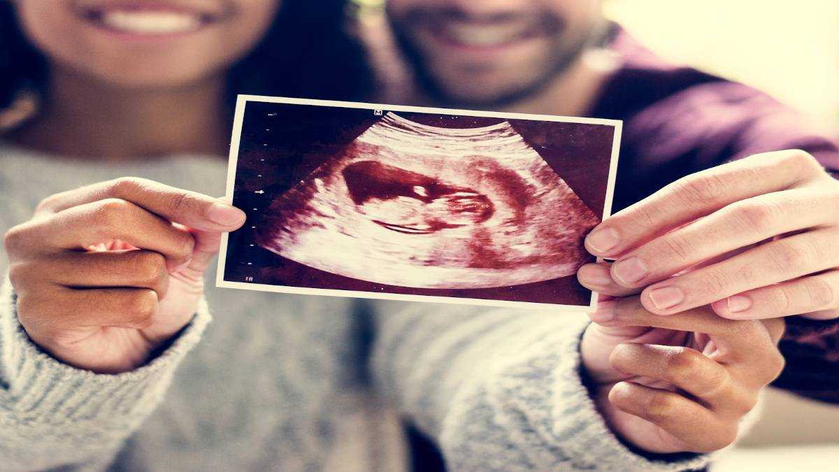
It not only helps in determining whether or not the fetus is growing properly but also helps the parents in connecting with their unborn. An ultrasound helps in confirming the heartbeat of the fetus, the baby’s anatomy, and general health.

In the first trimester –
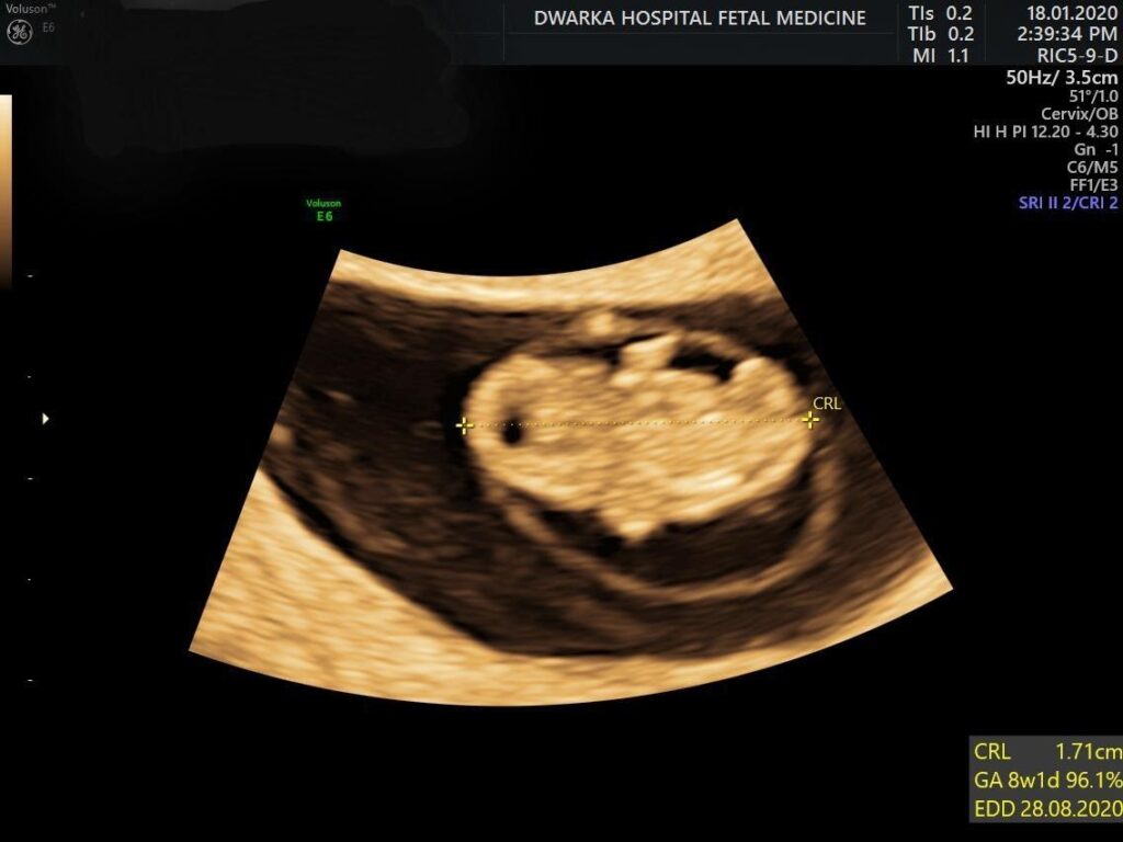
A. Dating Scan –
Between the sixth and the ten-week, an ultrasound is an inevitable part of the routine checkup. It is also called a dating scan. This ultrasound not only screens for the fetus’ heartbeat but also enables the parents to have a first glance at their baby.
An ultrasound performed in the first trimester of pregnancy helps in:
• Determining a due date – Measuring the fetus after the first trimester becomes difficult. Hence, in the first-trimester ultrasound, the fetus is effectively measured so as to determine a due date of delivery accurately.
• The heartbeat of the fetus is confirmed in the first-trimester ultrasound.
• The number of fetuses is determined. For women pregnant with twins, triplets, or such multiples, the first-trimester ultrasound is what helps determine the number of fetuses in the womb & their chorionicity.
• It also helps in ensuring that the pregnancy is on its normal course and that no abnormalities in the uterus and the fetus are noticed.
• While the first-trimester ultrasound is the most accurate in determining the gestational age, it also effectively measures the viability of a pregnancy. If pregnancy is showing any miscarriage symptoms, the doctor will suggest conducting ultrasound/s to check if everything is as supposed to be.
In the first trimester, doctors usually opt for a transvaginal ultrasound as opposed to an abdominal ultrasound. In this kind of an ultrasound, the doctor or his /her assistant inserts a probe in the vagina to measure the gestational sac, the yolk sac, the heart rate, and the fetal pole. In an abdominal scan, the bladder needs to be full while the doctor/assistant will apply a gel on the abdomen, and with the help of a transceiver, take measurements from different angles.
B. NT SCAN –
What is the NT scan, and how does it help?
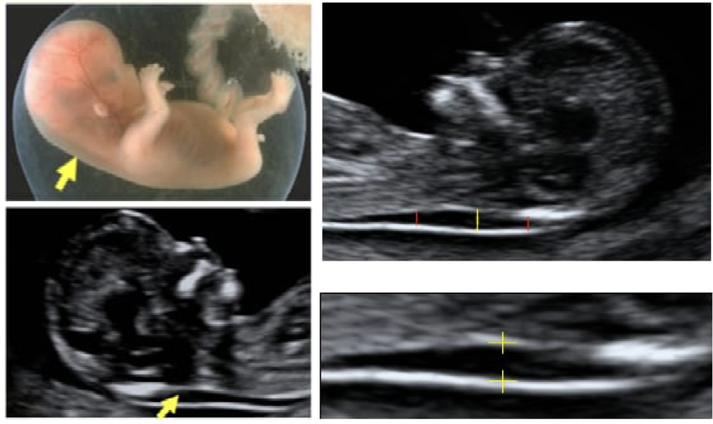
It is a common screening test that measures the size of a collection of fluid located at the back of the baby’s neck. This sonographic appearance of fluid under the skin behind the fetal neck in the first trimester of pregnancy is known as the nuchal translucency. The term translucency is used, irrespective of whether it is separated or not and whether it is confined to the neck or whole fetus. If it appears and its measurement is above 90% of fetal
CRL measurement, there is a possibility of the child having a chromosomal abnormality such as Down Syndrome, Edwards Syndrome, or Patau Syndrome. The NT scan is conducted by performing an abdominal, vaginal, or both ultrasound.
A nuchal translucency (NT) scan is conducted between the 11th and 13.6 weeks of pregnancy to check for any abnormalities in the fetus.
Combined screening is advised at the time of this scan for screening chromosomal abnormalities such as Down Syndrome, Edwards Syndrome, or Patau Syndrome.
If the ultrasound results are unclear in determining the health of the fetus or other details, the doctor will advise another ultrasound with a gap of a few days or weeks.
Special thanks to Dr. Sharad S Shinde (MBBS, DNB, FCPS, DGO, DGO) for the expert advice.